This is a procedure that involves inserting a fine telescope through the urethra (water-pipe) into the bladder and then up the ureter into the kidney. When the telescope is inserted only up to the ureter (eg for a stone in the ureter) it is called ureteroscopy. When the procedure involves inserting the telescope all the way up to the kidney, it is called renal pyeloscopy. Ureteropyeloscopy is a general term used to describe either of these operations.
The procedure can be performed using a straight instrument called a rigid ureteroscope, and this is done when access is only needed to the ureter. When access is needed into the kidney, a flexible instrument (flexible ureteroscope or flexible ureterorenoscope) is usually utilised.
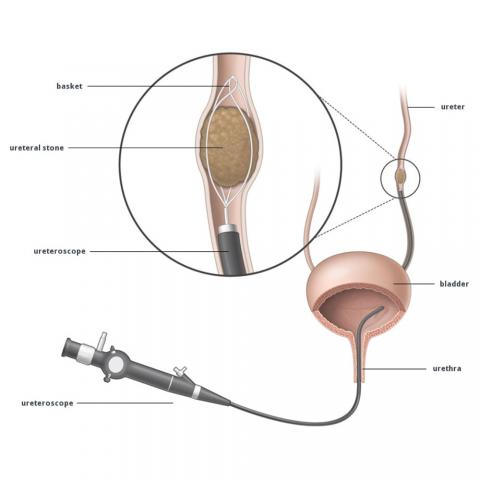
Ureteropyeloscopy is performed under a general anaesthetic, usually as a day case or an overnight stay in hospital. A ureteroscope is inserted into the ureter or kidney without the need for an incision. The stone is visualized directly and fragmented into small pieces using a Holmium Laser through a channel in the instrument. This type of laser will break up any stone – there is no stone which is too hard to fragment. The pieces of stone are then grasped with a basket and extracted out of the body, with only very fine pieces or powder left behind – these tiny fragments can easily pass naturally. The procedure usually takes 45 – 120 minutes with longer times required for larger stones.
The Ureteric Stent
Following stone extraction, a stent is usually left in the kidney. This is a hollow, fine soft tube which is placed between the kidney and bladder through the inside of the ureter. The stent allows free passage of urine from the kidney to the bladder without obstruction. A stent is needed after ureteropyeloscopy as the procedure results in bruising and swelling of the ureter that will cause ureter blockage if a stent is not placed.
The stent is usually removed after a few days to a few weeks depending on the particular circumstances. Ureteric stents cannot stay in the body indefinitely (unlike heart/coronary stents) as they will cause further stone formation leading to stent blockage. Removal of a stent is a minor and safe procedure which may be performed under local anaesthetic or with sedation as a day case in hospital. Stents not infrequently cause side effects – see Ureteric Stent.
Success Rate
There is little need for patients to be concerned about this procedure, particularly as Kidney Stone Melbourne Urologist Mr Uri Hanegbi is extremely experienced at treating kidney stones. Ureteropyeloscopy has a very high success rate at treating ureteric and renal stones, with much higher chances of success compared with Extracorporial Shockwave Lithotripsy (ESWL). Sometimes it is not possible to gain access to the stone with ureteropyeloscopy because the ureter is too narrow to fit the telescope (some people have very narrow ureters). In this circumstance, a stent is inserted into the kidney and the stone is not treated initially. The stent is much narrower than the telescope and it is nearly always possible to insert a stent even if the ureter is narrow. In this circumstance, the stent will gradually stretch the ureter over a period of a couple of weeks and at the time of stent removal, it is frequently much easier to insert the ureteroscope up to the stone in order to treat it.
In other words, ureteropyeloscopy nearly always requires two procedures. Usually, the first to treat the stone and the second to remove the stent; but at other times, to first insert a stent and then later to treat the stone at the time of stent removal. Patients with larger, more complex or difficult to access stones may even require a third procedure to achieve stone clearance.
Safety
Ureteropyeloscopy is a very safe procedure as no incision is performed and the body’s natural urinary channels are used to gain access to the stone. The risk of damaging the urinary system is very low. Ureter injury may occur if the ureter is tight or the stone very stuck in the ureter, but injuries to the ureter tend to heal themselves very well, without the need for surgical repair. Very rarely, a ureter may be damaged severely and this may require an open operation to repair the injury – this is extremely uncommon. Although the laser used to treat renal calculi is very powerful, it does not tend to damage the kidney as the laser only has a penetration of 0.4mm! Furthermore, the stone can be seen clearly and is fragmented under direct vision to make sure no other structures are damaged.
Side Effects
The side effects of ureteropyeloscopy tend to be mild. Loin pain is usually minor and many patients can therefore go home on the day of surgery. Visible blood in the urine is common and not a concern – this may continue until the stent is removed. Many patients will experience side effects from the presence of the ureteric stent. The risk of infection is low and the risk of damage to the urinary system is very low. Historically, there were reports of some patients developing a narrowing in their ureter following ureteroscopy due to formation of scar tissue - this is called a ureteric stricture. Now that surgical techniques and instruments have improved, this is a very rare complication.